Home
/ Knee Muscle Anatomy Mri : How To Read The Normal Knee Mri Kenhub, Cross sectional anatomy of the knee based on mri :
Knee Muscle Anatomy Mri : How To Read The Normal Knee Mri Kenhub, Cross sectional anatomy of the knee based on mri :
Knee Muscle Anatomy Mri : How To Read The Normal Knee Mri Kenhub, Cross sectional anatomy of the knee based on mri :. These muscles work in groups to flex, extend and stabilize the knee joint. These motions of the knee allow the body to perform such important movements as walking, running, kicking, and jumping. In conclusion, we describe the normal mri anatomy of the distal biceps femoris and the relationship of this muscle with the common peroneal nerve. Articular surface of patella and femur, condyle, epicondyle and muscles (popliteus, sartorius, gastrocnemius, semimembranous with tendos.) the images obtained were exported to jpeg from dicom data stored on the pacs (picture archiving and communicating system). Mri knee anatomy scroll using the mouse wheel or the arrows.
Assoc prof craig hacking and dr shu su et al. Cross sectional anatomy of the knee based on mri : It is the largest synovial joint in the body and allows flexion and extension of the leg as well as some rotation in the flexed position. T2w axial fat sat 1. Use the mouse scroll wheel to move the images up and down alternatively use the tiny arrows (>>) on both side of the image to move the images.
Mri Anatomy Of Knee Dr Muhammad Bin Zulfiqar from image.slidesharecdn.com These motions of the knee allow the body to perform such important movements as walking, running, kicking, and jumping. Doctors may recommend a knee mri if a patient experiences the following(3): Anatomy basic knee mri checklist. Use the mouse scroll wheel to move the images up and down alternatively use the tiny arrows (>>) on both side of the image to move the images.>>) on both side of the image to move the images. When a muscle has different orientations of the tendons it means that there are different patterns of edema possible depending on the tendon injured. The quadriceps muscles provide strength and power with knee extension (straightening). Anatomy basic knee mri checklist. The patellar tendon on the front of the knee is part of the quadriceps mechanism.
It is the largest synovial joint in the body and allows flexion and extension of the leg as well as some rotation in the flexed position.
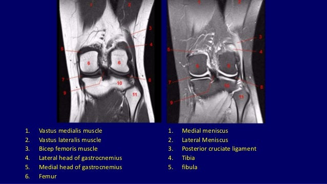
When a muscle has different orientations of the tendons it means that there are different patterns of edema possible depending on the tendon injured. Abnormal anatomy with normal signal, i.e. The images may also help physicians to distinguish normal, healthy tissues from dead tissues(2). It is the largest synovial joint in the body and allows flexion and extension of the leg as well as some rotation in the flexed position. Anatomy basic knee mri checklist. Doctors may recommend a knee mri if a patient experiences the following(3): The common peroneal nerve typically courses downward within abundant fat posterior to the short head of the biceps femoris muscle and superficial to the lateral head of the gastrocnemius muscle, but. In approximately 2% of the population, the anterior tibial artery branches along the keywords: Thigh muscles also protect neurovascular structures as they go through the proximal hip joint to the knee and lower leg (3). Use the mouse scroll wheel to move the images up and down alternatively use the tiny arrows (>>) on both side of the image to move the images. Coronal anatomy of the knee. Cross sectional anatomy of the knee based on mri : The muscles of the knee include the quadriceps, hamstrings, and the muscles of the calf.
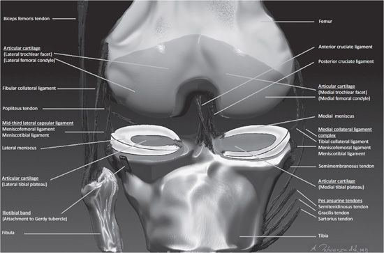
From superficial to deep includes the pes anserinus tendons, semimembranosus tendon, tibial collateral ligament, meniscofemoral and meniscotibial ligaments, and the medial meniscus. Anterior and posterior cruciate ligaments. This article is based on a presentation given by david rubin and adapted for the radiology assistant by robin smithuis. Coronal images are perpendicular to the long axis of the metatarsals. Assoc prof craig hacking and dr shu su et al.
Knee Mri Radiology Key from radiologykey.com Normal mr imaging anatomy of the knee. Assoc prof craig hacking and dr shu su et al. Anatomy basic knee mri checklist. Anatomy arthrogram anatomy basic shoulder mri. Naturally, in order to assess pathologic knee imaging, it is necessary to know the appearance of a normal knee mri. Use the mouse scroll wheel to move the images up and down alternatively use the tiny arrows (>>) on both side of the image to move the images. There is a flat area of tendon originating from the knee. Knee muscle anatomy axial mri :
Carefully labeled mris for all body parts, as well as schematic diagrams and concise statements, clarify correlations between bones and tissues.
Use the mouse scroll wheel to move the images up and down alternatively use the tiny arrows (>>) on both side of the image to move the images.>>) on both side of the image to move the images. Anatomy basic knee mri checklist. There is a flat area of tendon originating from the knee. Articular surface of patella and femur, condyle, epicondyle and muscles (popliteus, sartorius, gastrocnemius, semimembranous with tendos.) find this pin and more on anatomyby radiologist.ayman almatboly. From superficial to deep includes the pes anserinus tendons, semimembranosus tendon, tibial collateral ligament, meniscofemoral and meniscotibial ligaments, and the medial meniscus. The hamstrings muscles allow for strength and power in flexion (bending). Weak adductor muscles may cause knee instability and adductor strain (2). Anatomical structures of the lower limb (hip, thigh, knee, leg, ankle and foot) and specific regions (compartment of the lower. Coronal anatomy of the knee. The quadriceps muscles provide strength and power with knee extension (straightening). Use the mouse scroll wheel to move the images up and down alternatively use the tiny arrows (>>) on both side of the image to move the images. Atlas of knee mri anatomy. Mri wrist anatomy scroll using the mouse wheel or the arrows.
This article is based on a presentation given by david rubin and adapted for the radiology assistant by robin smithuis. Anatomy arthrogram anatomy basic shoulder mri. The patellar tendon on the front of the knee is part of the quadriceps mechanism. Normal mr imaging anatomy of the knee. Carefully labeled mris for all body parts, as well as schematic diagrams and concise statements, clarify correlations between bones and tissues.
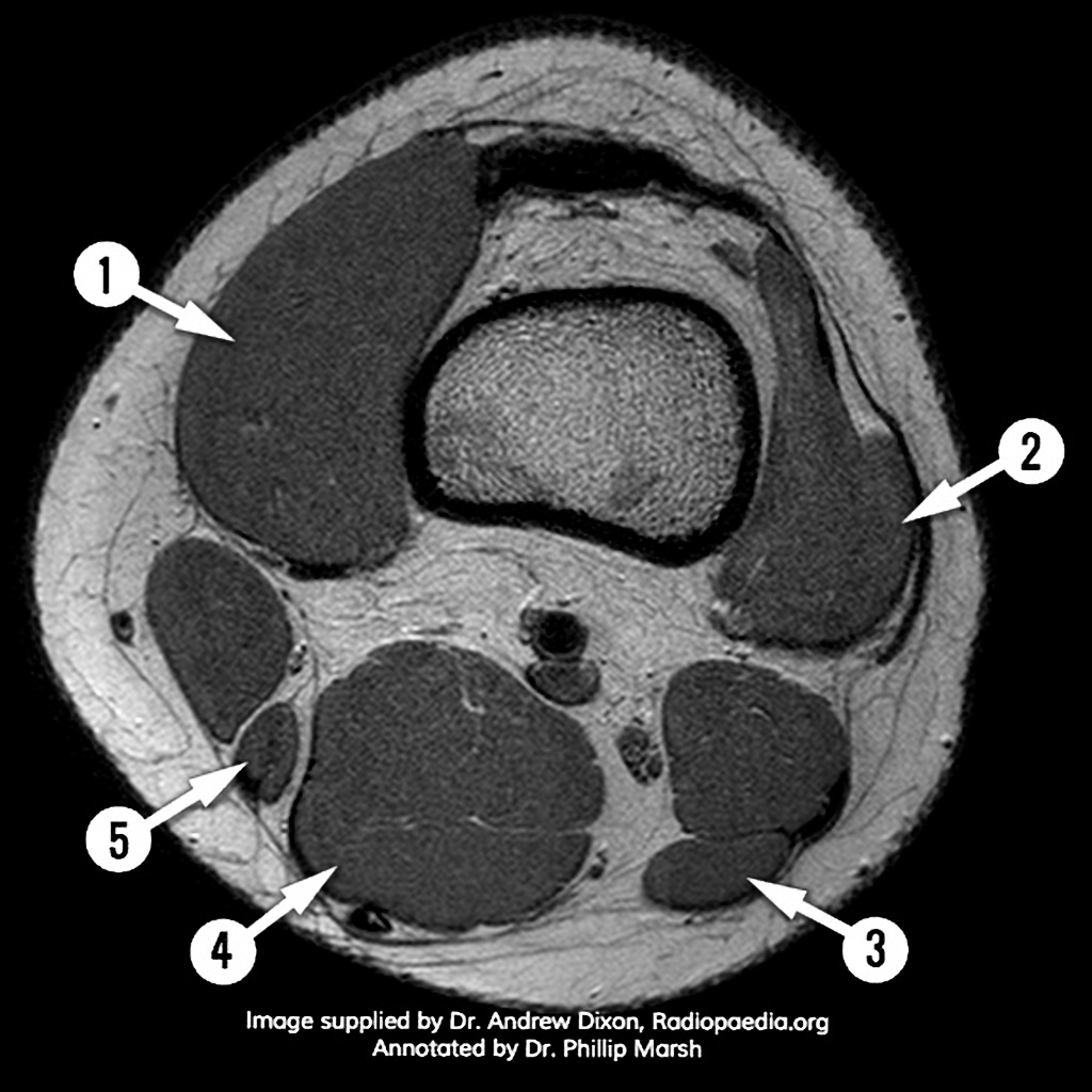
Mri Knee Axial Anatomy Quiz Radiology Case Radiopaedia Org from prod-images-static.radiopaedia.org Magnetic resonance imaging is particularly well suited for the medical evaluation of the musculoskeletal (msk) system including the knee, shoulder, ankle, wrist and elbow. The knee joint is a modified hinge joint between the femur tibia and patella. Anterior and posterior cruciate ligaments. There is a flat area of tendon originating from the knee. This mri hip joint axial cross sectional anatomy tool is absolutely free to use. Anatomy_of_knee_mri 7/16 anatomy of knee mri vessels, muscles, bone tendons, and ligaments facilitate accurate identification of key anatomic structures. It is the largest synovial joint in the body and allows flexion and extension of the leg as well as some rotation in the flexed position. From superficial to deep includes the pes anserinus tendons, semimembranosus tendon, tibial collateral ligament, meniscofemoral and meniscotibial ligaments, and the medial meniscus.
It is the largest synovial joint in the body and allows flexion and extension of the leg as well as some rotation in the flexed position.
Knee muscle anatomy axial mri : The hamstrings muscles allow for strength and power in flexion (bending). It is the largest synovial joint in the body and allows flexion and extension of the leg as well as some rotation in the flexed position. Doctors may recommend a knee mri if a patient experiences the following(3): Anatomy basic knee mri checklist. Abnormal anatomy with normal signal, i.e. In conclusion, we describe the normal mri anatomy of the distal biceps femoris and the relationship of this muscle with the common peroneal nerve. Cross sectional anatomy of the knee based on mri : Normal mr imaging anatomy of the knee. Magnetic resonance imaging mri is the imaging modality of choice in the diagnosis of acute and chronic soft tissue chondral and occult skeletal injuries of the knee. Carefully labeled mris for all body parts, as well as schematic diagrams and concise statements, clarify correlations between bones and tissues. This article is based on a presentation given by david rubin and adapted for the radiology assistant by robin smithuis. Articular surface of patella and femur, condyle, epicondyle and muscles (popliteus, sartorius, gastrocnemius, semimembranous with tendos.) the images obtained were exported to jpeg from dicom data stored on the pacs (picture archiving and communicating system).